Even today, few have heard of such a method of studying the body as scintigraphy. It is also called nuclear scanning. Despite this awesome name, the procedure is completely safe, painless and does not negatively affect the body.
Material Content:
What is the procedure for?
The name of this research method comes from the words scinti (lat.), Which means "to radiate light", and grapho - to "write." For the first time, radiopharmaceuticals in medicine were used at the beginning of the twentieth century, for which their creator was awarded the Nobel Prize. In the 60s, such methods began to develop intensively, then such studies as scanning and radiometry became famous. The development of a special camera became a new stage in the development of this kind of techniques. This method is called scintigraphy.
The procedure is carried out in order to identify various pathologies of the heart, lungs, brain, kidneys, bone system and other organs. Many of them are difficult to determine using other research methods. For example, osteoscintigraphy allows you to diagnose the disease 4-5 weeks earlier than a normal bone x-ray.
And also the procedure helps to clarify the degree of organ damage, the activity of the pathology in case of an already identified and diagnosed disease. In many cases, it allows you to track the effectiveness of the treatment.
What pathologies are identified during the study
Depending on the body being examined, the following pathologies can be identified during the procedure:
- various bone injuries (infectious or traumatic);
- Parkinson's disease;
- bone cancer;
- primary malignant neoplasms of the skeletal system - osteoma, osteosarcoma;
- heart defects;
- pathology of the circulatory system;
- malfunctions of the gallbladder - blockage of the ducts, pathological ejection of bile, stone formation;
- cysts, kidney tumors, the presence of stones or foci of infection;
- abscesses, cysts, spleen hematomas;
- pulmonary embolism;
- impaired liver function;
- Alzheimer's disease;
- epilepsy;
- multiple sclerosis;
- lymphomas
- stroke;
- thyroid adenoma.
The study helps to determine the nature of the spread of metastases in oncology and to predict the course of the disease.
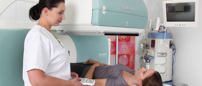
The principles of the apparatus
The principle of operation of the device is quite simple. Special gamma cameras take pictures of organs after they enter a radio indicator. A two-dimensional image is transmitted to the screen due to the emitted radiation. The substance is most often administered intravenously and enters the necessary organs, facilitating the research process.


Only in the case of diagnosis of pulmonary embolism does a person inhale a radio indicator, because that is how he quickly enters the lungs. The amount of radiopharmaceutical depends on the organ that needs to be examined. A large dose is needed to study the condition of the heart, brain, and bones, while a smaller dose is needed for other organs.
We can say that the radio indicator serves as a "transmitter" of information, since it emits gamma rays, which are recorded by a special camera.
The resulting images, or scintigrams, are divided into two categories:
- static - flat two-dimensional images. They are done in the study of the skeletal system, thyroid gland;
- dynamic - the result of combining several ordinary pictures. Allow to build dynamic curves. Used in the diagnosis and study of the functions of the liver, gall bladder, kidneys.
Tomographic and synchronized images are also distinguished. Doctors analyze the image data and then can make a specific diagnosis. Increased accumulation of a radio indicator in a diseased organ is called a “hot” focus, and its reduced or complete absence is called “cold”.
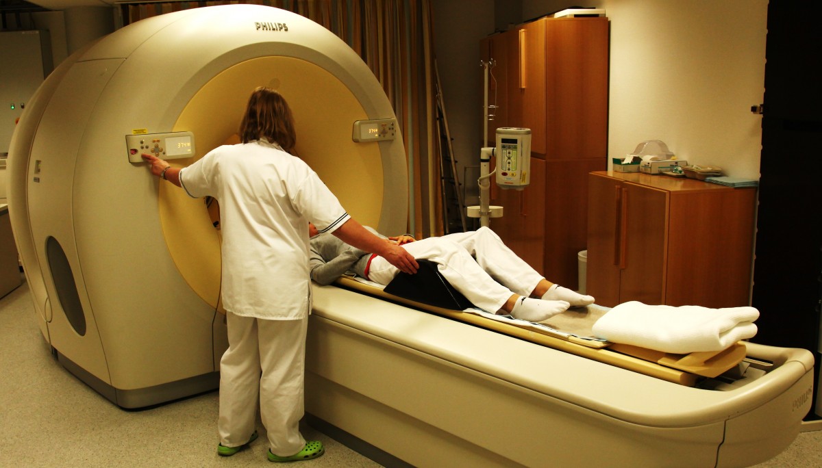
Scintigraphy process description
For the study of each organ, its own radio indicator is used. For example, for the diagnosis of thyroid diseases, this is a solution of pure technetium, heart pathologies - labeled tetrophosmin. The duration of the examination ranges from 25 minutes to 3.5 hours. It takes time for the active substance to reach a specific organ or tissue. For each organ, it is different. The bone tissue is examined the longest, the kidneys and thyroid gland are the least.
After the procedure, the substance introduced inside is excreted naturally during the day. There is no harm from radiopharmaceuticals, since they all have the property of continuous decay. The total radiation dose is much lower than with conventional x-rays.
Thyroid scintigraphy
This procedure takes a minimum of time - 20-25 minutes. She is appointed to assess the functioning of the body and its structure. The study helps to identify various pathological processes at an early stage.
Parathyroid scintigraphy should reveal a hormone-producing adenoma. In this case, the study time takes about 3 hours.
Skeleton bone examination
Skeletal bone scintigraphy is used to determine the exact cause of pain of unknown origin in the back, lower back and other parts of the body. The active substance is introduced inside and penetrates the skeletal system. This process is not quick, it can take several hours. After this, the cameras take pictures that allow you to diagnose the disease. Places where the radiopharmindicator has not penetrated or is very small are shown in the images as dark spots. It can talk about oncology.Bright spots often determine the presence of arthritis, fracture, or bone infection.
Special training is not required. But often doctors ask before the procedure to consume a lot of fluid, as this speeds up and facilitates the penetration of the indicator into the bones. After the introduction of the active substance, you need to wait a couple of hours until the specialist begins to take pictures. During this, you need to lie still, as otherwise the image will be blurry and fuzzy. The total study time is about 4 hours.
How is myocardial scintigraphy performed?
This procedure is necessary in order to better learn about the work of the heart. The study allows you to obtain a sufficient amount of information about the state of the heart muscle, the size of the chambers of the organ, and the movement of blood. This study is considered the leading diagnostic tool for coronary heart disease.
5 hours before the study, you must avoid drinking coffee and do not smoke. In rare cases, the doctor also recommends abstaining from food. The patient must notify the specialist if he is taking any medications, as certain medications can affect the result and distort it. The study takes 2-3 hours.
In some cases, the procedure is carried out under load: this means that in the interval between images the patient needs to do several physical exercises. This is necessary to see how the heart works in load mode.
Kidney analysis
Kidney scintigraphy helps to see firsthand how these organs work and to detect serious pathologies in time. Pictures allow not only to track blood flow, accurately determine the shape and size, but also to trace the process of formation and excretion of urine in each kidney. Bright spots may indicate an oncological tumor, dark spots - narrowing or blockage of blood vessels, cysts, scars, or infection.
Before this procedure, the patient is usually offered to drink 2-3 glasses of water to check how the kidneys function and how much time will pass before the indicator is displayed. The total duration of the procedure will be no more than 30 minutes.
To whom such a procedure is contraindicated
Although the procedure is considered safe, it is not recommended for pregnant women. In case of urgent need, nursing mothers can be scintigraphy, however, they can continue breastfeeding only after a day. During this time, drugs should be eliminated from the body.
And also research cannot be carried out for patients who are allergic to one of the elements of the introduced indicator.
Side effects are very rare. But sometimes patients may complain of sharp jumps in blood pressure and the associated malaise, frequent urination or allergic manifestations.
Often, a timely study helps to accurately diagnose and begin timely treatment. Therefore, if the doctor has prescribed scintigraphy for you, you should not put it off. Procrastination can be dangerous.


